Adhesive capsulitis of shoulder

Frozen shoulder, medically referred to as adhesive capsulitis, is a disorder in which the shoulder capsule, the connective tissue surrounding the glenohumeral joint of the shoulder, becomes inflamed and stiff, and grows together with abnormal bands of tissue, called adhesions, greatly restricting motion and causing chronic pain.
Adhesive capsulitis is a painful and disabling condition that often causes great frustration for patients and caregivers due to slow recovery. Movement of the shoulder is severely restricted. Pain is usually constant, worse at night, and along with the restricted movement can make even small tasks impossible. Certain movements can cause sudden onset of tremendous pain and cramping that can last several minutes.

This condition, for which an exact cause is unknown, can last from 5 months to 3 years or more, and is thought in some cases to be caused by injury or trauma to the area. It may also appear for no apparent reason. It is also believed that it may have an autoimmune component, with the body attacking healthy tissue in the shoulder. The condition may also cause chronic inflammation. Adhesions grow between the joints and tissue, greatly restricting motion and causing a number of painful complications. There is also a lack of fluid in the joint, further restricting movement.
In addition to difficulty with everyday tasks, people who suffer from adhesive capsulitis usually experience problems sleeping for extended periods due to pain that is worse at night and restricted movement/positions, resulting in chronic fatigue and other complications. The condition also can lead to depression, pain and problems in the neck and back, as well as damage to the tissue surrounding the area.
There are a number of risk factors for frozen shoulder, including diabetes, stroke, accidents, lung disease, connective tissue disorders, and heart disease. The condition very rarely appears in people under 40.
Treatment is painful and taxing, and consists of physical therapy, various medications, massage therapy, and in severe cases, surgery. A doctor may also perform manipulation under anesthesia, which breaks up the adhesions and scar tissue in the joint to help restore some range of motion.
In approximately 15% of patients, adhesive capsulitis may appear in both shoulders, at different times or together.

Presentation:

Movement of the shoulder is severely restricted. The condition is sometimes caused by injury that leads to lack of use due to pain but also often arises spontaneously with no obvious preceding trigger factor. These seemingly spontaneous cases are usually referred to as Idiopathic frozen shoulder. Rheumatic disease progression and recent shoulder surgery can also cause a pattern of pain and limitation similar to frozen shoulder. Intermittent periods of use may cause inflammation.
Abnormal bands of tissue (adhesions) grow between the joint surfaces, restricting motion. There is also a lack of synovial fluid, which normally helps the shoulder joint move by lubricating the gap between the humerus (upper arm bone) and the socket in the scapula (shoulder blade). It is this restricted space between the capsule and ball of the humerus that distinguishes adhesive capsulitis from a less complicated, painful, stiff shoulder. People with diabetes, stroke, lung disease, rheumatoid arthritis, and heart disease, or who have been in an accident, are at a higher risk for frozen shoulder. Adhesive capsulitis has been indicated as a possible adverse effect of some forms of highly active antiretroviral therapy (HAART). The condition rarely appears in people under 40 years old and (at least in its idiopathic form) is much more common in women than in men (70% of patients are women aged 40-60). Frozen shoulder in diabetic patients is generally thought to be a more troublesome condition than in the non-diabetic population. If a diabetic patient develops frozen shoulder then the time to full recovery is often prolonged from the usual 12 month period. Cases have also been reported after breast or lung surgery.

Prevention:

To prevent the problem, a common recommendation is to keep the shoulder joint fully moving to prevent a frozen shoulder. Often a shoulder will hurt when it begins to freeze. Because pain discourages movement, further development of adhesions that restrict movement will occur unless the joint continues to move full range in all directions (adduction, abduction, flexion, rotation, and extension). Therapy will help one continue movement to discourage freezing and warm it. A medical doctor referral is needed before? physical therapy.

Signs and diagnosis:

With a frozen shoulder, one sign is that the joint becomes so tight and stiff that it is nearly impossible to carry out simple movements, such as raising the arm. People complain that the stiffness and pain worsen at night. Pain due to frozen shoulder is usually dull or aching. It can be worsened with attempted motion. A doctor, or therapist (occupational, massage or physical), may suspect the patient has a frozen shoulder if a physical examination reveals limited shoulder movement.
Frozen shoulder can also be diagnosed if limits to the active range of motion (range of motion from active use of muscles) are the same or almost the same as the limits to the passive range of motion (range of motion from a person manipulating the arm and shoulder). An arthrogram or an MRI scan may confirm the diagnosis – although in practice this is rarely required. Most orthopaedic specialists make the diagnosis of frozen shoulder by recognising the typical pattern of signs and symptoms.

Physicians have described the normal course of a frozen shoulder as having three stages:

  • Stage one: The “freezing” or painful stage, which may last from six weeks to nine months, and in which the patient has a slow onset of pain. As the pain worsens, the shoulder loses motion.
  • Stage two: The “frozen” or adhesive stage is marked by a slow improvement in pain, but the stiffness remains. This stage generally lasts four months to nine months.
  • Stage three: The “thawing” or recovery, during which shoulder motion slowly returns toward normal. This generally lasts five months to 26 months.
Management:

Management of this disorder focuses on restoring joint movement and reducing shoulder pain. Usually, it begins with nonsteroidal anti-inflammatory drugs (NSAIDs).
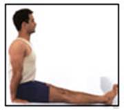
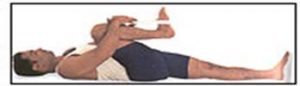
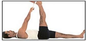
Physiotherapy treatments can help the situation. The treatment include interferential therapy, ultrasound, heat or cold therapy. Follow by gentle passive and active stretching exercises. These stretching exercises, which may be performed in the home with the help of a physiotherapist , are the treatment of choice. Treatment may be needed for several months.
If these measures are unsuccessful, the doctor may recommend manipulation of the shoulder under general anesthesia to break up the adhesions. Surgery to cut the adhesions is only necessary in some cases. Surgery to correct other problems with the shoulder may also be needed.

Tennis Elbow

Tendonitis of the elbow is often referred to as tennis elbow due to its common occurrence in tennis players, but in fact any sport or activity that requires gripping can cause this problem.
A more precise definition of Tennis elbow is that it is an inflammation of several structures of the elbow. These include muscles, tendons, bursa, periosteum, and epicondyle (bony projections on the outside and inside of the elbow, where muscles of the forearm attach to the bone of the upper arm). This condition is also called epicondylitis, lateral epicondylitis, medial epicondylitis, or golfer’s elbow, where pain is present at the inside epicondyle. Such pain is usually more noticeable during or after stressful use of the arm.
In sever cases, lifting and grasping even light things may be very painful.

Causes:

Tennis elbow occurs when their is damage to the muscles, tendons and ligaments around the elbow joint and forearm. Small tears, called micro tears, form in the tendons and muscles, which control the movement of the forearm. They cause a restriction of movement, inflammation and pain. These micro tears eventually lead to the formation of scar tissue and calcium deposits.
If untreated, this scar tissue and calcium deposits can put so much pressure on the muscles and nerves that they can cut off the blood flow and pinch the nerves responsible for controlling the muscles in the forearm. Often this happens during a backhand stroke in racket sports, especially if the stroke technique is poor. Another cause is overuse of the muscles in a repetitive way. Examples of this are prolonged use of a screwdriver or typing.

Prevention:

Obviously prevention is better than cure so I will first discuss a few prevention options:
A thorough warm up will help to prepare the muscles and tendons for any activity to come. Without a proper warm up the muscles and tendons will be tight and stiff. There will be limited blood flow to the forearm area, which will result in a lack of oxygen and nutrients for the muscles. This is a definite recipe for a muscle/ tendon injury.
Having flexible muscles and tendons is extremely important in the prevention of most strain or sprain injuries. When muscles and tendons are flexible and supple, they are able to move and perform without being over stretched. If however, your muscles and tendons are tight and stiff, it is quite easy for those muscles and tendons to be pushed beyond their natural range of movement. When this happens strains, sprains, and pulled muscles occur.

Treatment:

Treatment may include stopping or limiting activities that cause the pain, such as heavy lifting with the palm facing down. Sometimes a band wrapped around the forearm near the elbow is used to protect the injured muscles as they are healing. In some cases, the wearing of a wrist splint may be recommended for the same purpose.
Non-steroidal anti-inflammatory drugs (NSAIDs), such as naproxen or ibuprofen may be given for pain. Injections of cortisone may also be used to relieve the pain.
Physiotherapy treatment, such as interferential therapy, ultrasound, laser therapy, heat or cold treatment to promote the healing process. A tennis elbow band may be advised. Your physiotherapist may recommend exercises that stretch and strengthen the muscles to help prevent the condition from returning. Some patients respond to additional treatment through therapy. As the condition improves, there is usually a slow return to normal activities. Recurrence of this condition is common.

Osteoarthritis of Knee

Osteoarthritis is a type of arthritis that is caused by the breakdown and eventual loss of the cartilage of one or more joints. Cartilage is a protein substance that serves as a “cushion” between the bones of the joints. Osteoarthritis is also known as degenerative arthritis. Osteoarthritis commonly affects on joints as large weight-bearing joints, such as the hips and knees. Most cases of osteoarthritis have no known cause and are referred to as primary osteoarthritis. When the cause of the osteoarthritis is known, the condition is referred to as secondary osteoarthritis.

Causes:

Primary osteoarthritis is mostly related to aging. With aging, the water content of the cartilage increases and the protein makeup of cartilage degenerates. Repetitive use of the joints over the years irritates and inflames the cartilage, causing joint pain and swelling. Eventually, cartilage begins to degenerate by flaking or forming tiny crevasses. In advanced cases, there is a total loss of the cartilage cushion between the bones of the joints. Loss of cartilage cushion causes friction between the bones, leading to pain and limitation of joint mobility. Inflammation of the cartilage can also stimulate new bone outgrowths (spurs) to form around the joints. Osteoarthritis occasionally can be found in multiple members of the same family, implying an heredity (genetic) basis for this condition.

Secondary osteoarthritis is caused by another disease or condition. Conditions that can lead to secondary osteoarthritis include obesity, repeated trauma or surgery to the joint structures, abnormal joints at birth (congenital abnormalities), gout, diabetes and other hormone disorders. Obesity causes osteoarthritis by increasing the mechanical stress on the cartilage. In fact, next to aging, obesity is the most powerful risk factor for osteoarthritis of the knees. The early development of osteoarthritis of the knees among weight lifters is believed to be in part due to their high body weight. Repeated trauma to joint tissues (ligaments, bones and cartilage) is believed to lead to early osteoarthritis of the knees in soccer players. Interestingly, recent studies have not found an increased risk of osteoarthritis in long-distance runners.
Crystal deposits in the cartilage can cause cartilage degeneration, and osteoarthritis. Uric acid crystals cause arthritis in gout, while calcium pyrophosphate crystals cause arthritis in pseudogout. Some people are born with abnormally formed joints (congenital abnormalities) that are vulnerable to mechanical wear, causing early degeneration and loss of joint cartilage. Osteoarthritis of the hip joints is commonly related to design abnormalities of these joints that had been present since birth. Hormone disturbances, such as diabetes and growth hormone disorders, are also associated with early cartilage wear and secondary osteoarthritis.

Symptoms:

The most common symptom of osteoarthritis knee is pain in the knee joint after repetitive use. Joint pain is usually worse later in the day. There can be swelling, warmth, and creaking of the affected joints. Pain and stiffness of the joints can also occur after long periods of inactivity, for example, sitting in a heater. In severe osteoarthritis, complete loss of cartilage cushion causes friction between bones, causing pain at rest or pain with limited motion. Patient may have difficulty in walking up and down stairs. Even worse, walking ground level may have difficulty.

Treatment:

Aside from weight reduction and avoiding activities that exert excessive stress on the joint cartilage, there is no specific treatment to halt cartilage degeneration or to repair damaged cartilage in osteoarthritis. The goal of treatment in osteoarthritis is to reduce joint pain and inflammation while improving and maintaining joint function.
Some patients with osteoarthritis have minimal or no pain, and may not need treatment. Others may benefit from conservative measures such as rest, exercise, weight reduction and physiotherapy, and mechanical support devices.
In fact, even modest weight reduction can help to decrease symptoms of osteoarthritis of the large joints. Medications are used to complement the physical measures described above. Medication may be used topically, taken orally, or injected into the joints to decrease joint inflammation and pain. When conservative measures fail to control pain and improve joint function, surgery can be considered.
Resting sore joints decreases stress on the joints, and relieves pain and swelling. Patients are asked to simply decrease the intensity and/or frequency of the activities that consistently cause joint pain. Exercise usually does not aggravate osteoarthritis when performed at levels that do not cause joint pain. Exercise is helpful in osteoarthritis in several ways. First, it strengthens the muscular support around the joints. It also prevents the joints from “freezing up” and improves and maintains joint mobility. Finally, it helps with weight reduction and promotes endurance. Applying local heat before and cold packs after exercise can help relieve pain and inflammation. Swimming is particularly suited for patients with osteoarthritis because it allows patients to exercise with minimal impact stress to the joints. Other popular exercises include walking, stationary cycling, and light weight training.
Physiotherapy can provide to decrease the inflammation of knee joint as well as promote healing, such as interferential therapy, ultrasound, laser, heat and cold therapy. Also, strengthening exercises and mobilization exercises are important to help prevent recurrence of pain. They may also be help by supportive devices, such as splints, canes, walkers, and braces. These devices can be helpful in reducing stress on the joints. Treatments may last few weeks to months for better improvement.

Sciatica

The diagnosis of ‘sciatica’ means that there is inflammation of the sciatic nerve. The sciatic nerve supplies information about movements to the leg, and sends information about sensations back to the brain. The sciatic nerve is quite large; in fact, it is the largest peripheral nerve in the body.
The sciatic nerve is formed from the lower segments of the spinal cord; it is made up from the lumbar and sacral nerve roots from the spine. The sciatic nerve exits the lower part of the spinal cord (lumbosacral region), passes behind the hip joint, and runs down the back of the thigh.

How does this nerve normally function?

The sciatic nerve, like most other nerves, performs two basic functions: first, it sends signals to your muscles from the brain; and second, it collects sensory information from the legs and passes this back to your brain. Conditions such as sciatica that affect the nerve will alter these normal functions. This usually manifests as either weakness of these leg muscles, pain in the legs and thighs, or both.

What happens to cause sciatica?

The most common cause of sciatica is a herniated spinal disc. When this happens, the normal cushion between the vertebra of your spine ruptures. This causes the disc to push out into areas normally occupied by these nerves. The nerves are compressed and people then experience the symptoms of pain, weakness, and numbness. Other conditions, such as spinal stenosis, spondylolisthesis, or piriformis syndrome can also cause cause sciatica symptoms by irritating the nerve.

What are the signs and symptoms of sciatica?

As stated above, sciatica can cause both sensory and muscular abnormalities in the legs and thighs. Common symptoms of sciatica include:

  • A cramping sensation of the thigh
  • Shooting pains from the buttock, down the leg
  • Tingling, or pins-and-needles sensations in the legs and thighs
  • A burning sensation in the thigh

In addition, patients with sciatica may notice a worsening of their symptoms with maneuvers such as squatting or coughing. These maneuvers can increase pressure around the nerve and magnify the symptoms of sciatica.

Who is prone to developing symptoms of sciatica?

Sciatica can affect just about anyone, but it is extremely uncommon in young patients. Sciatica typically affects 30 to 50 year old patients. Often there is a sudden onset that may be attributed to over-exertion or a back injury.

What do I need to do for sciatica?

Most importantly, you need to find out if sciatica is the cause of your symptoms. Your doctor will take a thorough history, perform a physical exam, and test several specific functions of the nerve. Several other conditions may cause hip and thigh pain, and need to be considered. It is important to determine the correct cause of your symptoms prior to beginning treatment of sciatica. Other tests, including X-Rays or possibly an MRI may be helpful, but they may not needed.

What treatments are available for sciatica?

Exercises and physical therapy are helpful. Many people find that heat packs and ice packs soothe the muscles that are painful in sciatica. Some doctors may prescribe an epidural steroid injection that can deliver anti-inflammatory medication directly to the inflamed area around the nerves.
Surgical treatment of sciatica is not usually needed, but in individuals who undergo the above treatments for a minimum of three months, and still have symptoms, surgery may be considered. The surgical procedure is one that allows more room for the nerve in the area being compressed. This may mean removing the ruptured disc, opening up the bone around the nerve, or a combination of both.

Will I get better from sciatica?

This is the good news. Most people (80-90%) fully recover from sciatica without surgery. In most cases the nerve is not permanently damaged, and individuals recover in the 3-week to 3-month time frame.
Sciatica is not a medical emergency. However, if you experience difficulty with bowel or bladder function, decreased sensation around the genitals, or progressive leg weakness, this may be the sign of cauda equina syndrome, a medical emergency. If you have these symptoms, contact your doctor or go to the emergency room immediately.

De Quervain’s disease


De Quervain’s disease is a painful inflammation of tendons in the thumb that extend to the wrist (tenosynovitis). The swollen tendons and their coverings rub against the narrow tunnel through which they pass. The result is pain at the base of the thumb and extending into the lower arm.

What Causes de Quervain’s Disease?

Often, the cause of de Quervain’s disease is unknown, but overuse, a direct blow to the thumb, repetitive grasping, and certain inflammatory conditions such as rheumatoid arthritis can all trigger the disease. Gardening, racquet sports, and various workplace tasks may also aggravate the condition.

Who Gets de Quervain’s Disease?

While anyone can get de Quervain’s, it affects women eight to 10 times more often than men. Oddly, it may occur in the time just after pregnancy

What Are the Symptoms of de Quervain’s Disease?

Pain along the back of the thumb, directly over two thumb tendons, is common in de Quervain’s. The condition can occur gradually or suddenly; in either case, the pain may travel into the thumb or up the forearm. Thumb motion may be difficult and painful, particularly when pinching or grasping objects. Some people also experience swelling and pain on the side of the wrist at the base of the thumb. The pain may increase with thumb and wrist motion. Some people feel pain if direct pressure is applied to the area.

How Is de Quervain’s Disease Diagnosed?

The test most frequently used to diagnose de Quervain’s disease is the Finkelstein test. Your doctor will ask you to make a fist with your thumb placed in your palm. When the wrist is bent toward the outside, the swollen tendons are pulled through the tight space and stretched. If this movement is painful, you may have de Quervain’s disease.

How Is de Quervain’s Disease Treated?

Ice may be applied to reduce inflammation. If symptoms continue, your doctor may give you anti-inflammatory medication such as naproxen or ibuprofen or may inject the area with steroids to decrease pain and swelling. Normally, your medical doctor would refer you to visit physiotherapist to promote the healing and rehabitation.

Physiotherapy

Physitherapist would use proper treatment method according to your examination.

Electrotherapy

Utilize magnetic therapy, laser, mircocurrent, short wave, interfertial therapy, ultrasound, acupuncture and shock wave to promote circulation which can decrease inflammation and pain.

Assistive Devices

using proper wrist protector and Taping can reduce inflammation and pain which can promote the healing process.

Exercise Therapy

The following exercises need to according to your physiotherapist instrction

  1. Thumb stretching exercise
    Using 4 fingers to warp around the thumb, slightly unlar deviated (pointing toward the ground) until feel slightly tight, hold it for 15-20 seconds, relax slowly and repeat 5-10 times. Perform 2-3 sets per day.
  2. fingers strengthening exercise
    Use a rubber band to wrap around thumb, second and ring finger with palm facing up, open your fingers and hold it for 10 seconds. Repeat 3-5 minutes and perform it 3 sets per day.
Note

  1. increase the symptom when holding baby inproperly
    Using your wrist and thumb to hold baby’s buttock area would increase the stress of shoulder and wrist region.
  2. Using forearm instead of using wrist region to hold the buttock area of baby would decrease the stress on shoulder. Remind yourself to press down your shoulder region when holding a baby.